BioTek Lionheart FX Automated Microscope
RUO
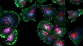
Lionheart FX automated microscope is a compact system for a broad range of imaging workflows. It offers up to 60x and 100x oil immersion magnification, with fluorescence, brightfield, color brightfield, and phase contrast channels for maximum application reach.
An optional environment control cover provides incubation to 40 °C and effective containment for CO2/O2 control. A humidity chamber optimizes conditions for long-term live cell imaging applications, and an available dual reagent injector facilitates rapid kinetic assays.
For Research Use Only. Not for use in diagnostic procedures.
- Automated Cell Imagers
Request a Quote
Download the product brochure using the DOWNLOADS button above, or
Product Details
Features
- Small footprint for easy installation on most standard lab benches
- No external instrumentation or dedicated darkroom required for image capture
- Wide field of view (WFOV) camera provides fast automated imaging in microplates and slides, enabling researchers to get the answers they need quickly
- Gen5 imaging software offers unique, full automation to capture, process, analyze, and publish workflows
- Environmental controls enable Lionheart FX to image and analyze live cell assays over time
- Laser-based and software-based autofocus capture sharp, in-focus images in automated microscopes
- Open design of the Lionheart FX stage allows researchers to work with microfluidic devices
- Dual reagent injector module for Lionheart FX allows fast cellular reactions to be imaged after the addition of a reagent
Specifications
Environment Control |
|
Imager/Reader Options |
|
Imaging Methods |
|
Imaging Modes |
|
Magnification Range |
|
Microscope Type |
|
Supported Labware |
|
How It Works
Applications
Technology
Proprietary Laser Autofocus
Laser autofocus and image-based (or software-based) autofocus are two methods of autofocus widely used in microscopy. Learn MoreCell Counting
Accurate cell counting is a critical step across a spectrum of cell biology applications. Direct cell counting uses methods that identify and enumerate individual cells, as opposed to indirect methods that estimate population sizes using biochemical methods. Learn MoreObject Tracking
With the incorporation of the object tracking module, available within Agilent BioTek Gen5 software for imaging and microscopy, this can be easily accomplished in a fully automated fashion. Learn MoreAutomatic Region of Interest Identification
This process enables walk-away slide scanning for four or more slides, if combined with Agilent automation offerings, and can be used with the Agilent BioTek Lionheart automated microscopes and Agilent BioTek Cytation imaging multimode readers. Learn MoreSpot Counting
The spot counting module available in Agilent BioTek Gen5 software enables the number of spots counted, and other information, to be tied to the original primary mask data, allowing these values to be known for each object initially identified in the image. Learn More4-Zone Incubation and Condensation Control
Many Agilent single and multi-mode microplate readers include an incubation function for temperature sensitive applications, such as kinetic assays. Some microplate reader incubators use a heating block with a fan to circulate the hot air and warm the reading chamber and microplate. Learn MoreAugmented Microscopy
Agilent BioTek instrumentation and software create the unique augmented microscopy experience with the integration and automation of all steps from image capture to publication-ready data. Learn MoreLiterature
- Key Literature
-
Download Literature
Download a product brochure or data sheet- Brochures
- English
- 23 Nov 2021
- 72.95 KB
- Application Notes
- Posters
- Specifications
- Technical Overviews
Support
Didn’t find what you need? Contact our dedicated Cell Analysis support teams.
Tools
Explore customer reviews of this product or write your own review
Image Gallery
Click here to view our Image Gallery, including images captured with Agilent BioTek instruments.Agilent Cell Analysis Publication Database
Search scientific publications by research area, cell type and more.Videos
Training & Events
Tools and Techniques for Optimizing Cell Proliferation Studies
In this webinar we provide a detailed review of available methods for measuring cell proliferation, with a particular focus on recent assay innovations, including both 2D and 3D formats.
Webinar/Training
3D Culture Methods and Imaging Considerations
We cover the advantages and limitations of the various culture models, assay formats, and imaging approaches.
Webinar/Training
A Practical Guide for 3D Cell Culture Systems and Optimizing Spheroid Imaging Assay - Part 1 of 2
3D cell culture systems have become a well-established in vitro experimental approach to model human disease for a broad range of research applications.
Webinar/Training
Related Products
Promotions